Identification of Enriched PTM Crosstalk Motifs from Large-Scale Experimental Data Sets

ABSTRACT: Post-translational modifications (PTMs) play an impor-
tant role in the regulation of protein function. Mass spectrometry based
proteomics experiments nowadays identify tens of thousands of PTMs in
a single experiment. A wealth of data has therefore become publically
available. Evidently the biological function of each PTM is the key
question to be addressed; however, such analyses focus primarily on
single PTM events. This ignores the fact that PTMs may act in concert
in the regulation of protein function, a process termed PTM crosstalk.
Relatively little is known on the frequency and functional relevance of
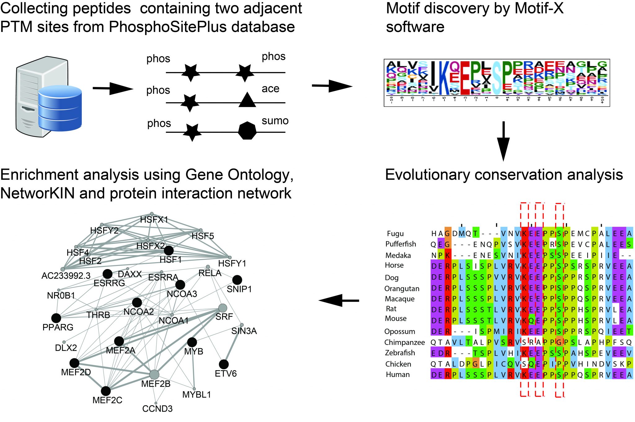
crosstalk between PTM sites. In a bioinformatics approach, we extracted
PTMs occurring in proximity in the protein sequence from publically
available databases. These PTMs and their flanking sequences were
subjected to stringent motif searches, including a scoring for evolutionary
conservation. Our unprejudiced approach was able to detect a respectable set of motifs, of which about half were described previously. Among these we could add many new proteins harboring these motifs. We extracted also several novel motifs, which through their widespread appearance and high conservation may pinpoint at previously nonannotated concerted PTM actions. By employing network analyses on these proteins, we propose putative functional roles for these novel motifs with two PTM sites in close proximity.


Publication: Defining the background in protein network analysis; the CRAPome

Albert Heck, Tech Yew Low, Vincent Halim and Shabaz Mohammed of the Biomolecular Mass Spectrometry and Proteomics Group (Utrecht University) joined an international effort involving many renowned proteomics researchers and cell biologists to contribute to a contaminant repository for protein affinity purifications termed the CRAPome. The CRAPome aims to create a compendium for all background contaminants derived from negative controls of AP-MS (affinity purification-MS) experiments based on various categories such as epitope tags, beads, cell lines or organisms and experimental protocols used in different labs. Although AP-MS is widely used for identifying protein-protein interactions, discriminating bona fide interactors from the background contaminants is often a daunting task. In this work, we show that, by aggregating negative controls from multiple AP-MS studies, we increase coverage and improve the characterization of background associated with a given AP-MS protocol. The current version of the CRAPome comes with online toolkits, which compute scoring functions to guide research community in decision-making. All researchers are also encouraged to contribute their negative controls to the CRAPome so as to make it an even more comprehensive resource. The CRAPome is freely available at http://www.crapome.org/.

CRAPome

The associated article was published in Nature Methods 7th of July 2013 (http://www.nature.com/nmeth/journal/vaop/ncurrent/full/nmeth.2557.html).

Fighting the cause of Chagas disease

Reducing the parasite population with a virus

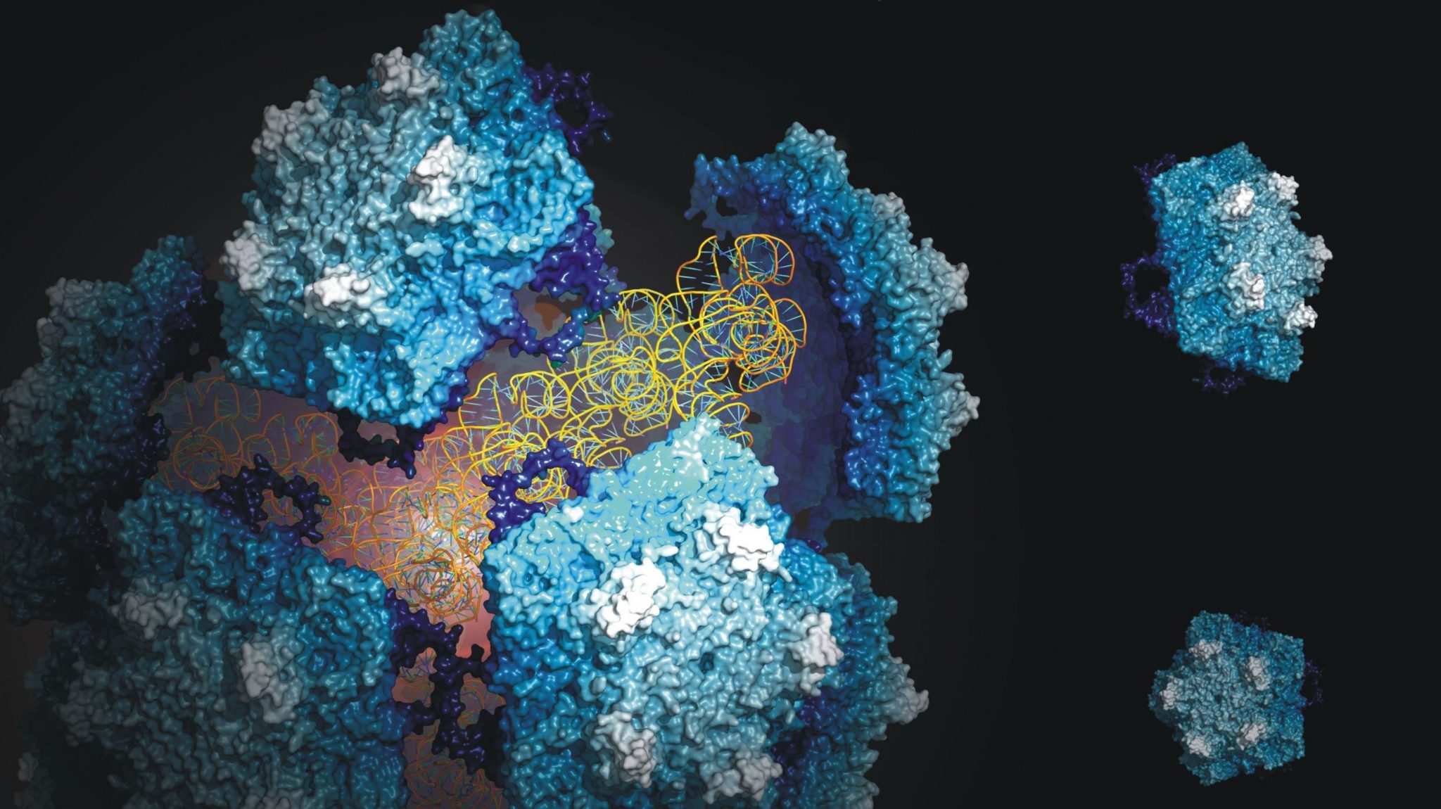
The hitherto fairly unknown Triatoma virus, which kills insects that spread the tropical disease known as Chagas disease, could be used as a pesticide to control the insect plague and thus the spread of the disease. Researchers of the Biomolecular Mass Spectrometry and Proteomics Group in collaboration with researchers of VU University Amsterdam have studied every little detail of the virus and published their findings on 28 April in Nature Chemistry.

 According to assessments of the World Health Organisation, ten million people had Chagas disease in 2012. At least 20,000 people die from the disease every year. This tropical disease is transferred by parasites in blood-sucking insects. The disease can be treated in the first few months, but when a longer period of time has elapsed, patients can no longer be saved. The disease is prevalent in Latin America, but has meanwhile spread to the north and infections have been found in the North-American state of Florida.

Fighting the cause
One of the best ways to prevent the spread of the disease is to combat the insect population that carries the pathogenic parasite. The Triatoma virus offers options in this respect, because infection with the virus kills the insects. In an attempt to chart this virus, researchers of the group of Professor Albert Heck (Utrecht University) in cooperation with the group of Dr Wouter Roos (VU University Amsterdam) have studied the composition, structure and stability of the virus in detail with the help of mass spectrometry and atomic force microscopy.

A complex collaboration
The researchers have shown that the stability of the virus is determined by a complex collaboration between the virus and its genetic material, which is strongly influenced by environmental factors. The experiments also provided information about the structure of the virus and the functioning of the genome. Both processes are vital to the virus’ life cycle. Furthermore, the data contributes to the design of viral systems in nano-technology and medicine.

Structure and stability
Albert Heck: “Our work has provided a lot of insight into the structure and stability of the virus. We have used many techniques in order to find out how to extract the genome from the virus and to reassemble the virus afterwards. This offers opportunities to add new elements to the virus which are potentially lethal to the host.”


This research was sponsored by the Netherlands Proteomics Centre, the Netherlands Organisation for Scientific Research (NWO) and the Foundation for Fundamental Research on Matter (FOM)

Publication
J. Snijder, C. Uetrecht, R.J. Rose, R. Sanchez-Eugenia, G.A. Marti, J. Agirre, D.M.A. Guerin, G.J.L. Wuite, A.J.R. Heck, W.H. Roos
Probing the biophysical interplay between a viral genome and its capsid.
Nature Chemistry

 

Top publication: Deletion of genes causes cancer

In a recent Nature paper researchers from the Hubrecht Institute in cooperation with researchers from the University Medical Center Utrecht (UMC Utrecht), University Utrecht (UU) and the Netherlands Proteomics Centre (NPC), describe a gene that limits the growth of intestinal adenomas. The research might be a lead in the treatment of intestinal cancer. Stem cells in the gut continuously provide new tissue. Prof. Hans Clevers succeeded earlier in identifying and isolating these stem cells.

Together with Dr. Madelon Maurice of the UMC Utrecht and Prof. Albert Heck of the UU and the NPC, the Clevers group searched for genes which are only active in the intestinal stem cells. They found RNF43. When this gene is deleted, exponential growth of the intestinal stem cells cause adenomas, a pre stage of intestinal cancer.

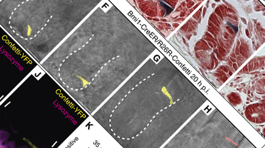
LGR5+ stem cells reside at crypt bottoms, intermingled with Paneth cells that provide Wnt, Notch and epidermal growth factor signals. In this article the researchers find that the related RNF43 and ZNRF3 transmembrane E3 ubiquitin ligases are uniquely expressed in LGR5+ stem cells. Simultaneous deletion of the two genes encoding these proteins in the intestinal epithelium of mice induces rapidly growing adenomas containing high numbers of Paneth and LGR5+ stem cells. In vitro, growth of organoids derived from these adenomas is arrested when Wnt secretion is inhibited, indicating a dependence of the adenoma stem cells on Wnt produced by adenoma Paneth cells. In the HEK293T human cancer cell line, expression of RNF43 blocks Wnt responses and targets surface-expressed frizzled receptors to lysosomes. In the RNF43-mutant colorectal cancer cell line HCT116, reconstitution of RNF43 expression removes its response to exogenous Wnt. We conclude that RNF43 and ZNRF3 reduce Wnt signals by selectively ubiquitinating frizzled receptors, thereby targeting these Wnt receptors for degradation.

Top Publication: Genes & proteins involved in the regeneration of the small intestine uncovered

Genes & proteins involved in the regeneration of the small intestine uncovered

In a collaboration involving several partners in the Utrecht Life Sciences Initiative (ULS) members of the Netherlands Proteomics Centre (Heck group) and the Hubrecht Institute (Clevers group, Alexander van Oudenaarden group, presently still at the Department of Physics, MIT, Cambridge, USA) made a significant advance in understanding the molecular basis that program the regeneration of tissue through the stem cells in the intestine. Joint efforts from these groups allowed the identification of genes and proteins that are specifically expressed in the stem cells of the intestine and that allow this tissue to regenerate. These results have been jointly published in The EMBO Journal on June 12, 2012.

Stem cells receive special attention in the scientific community and in society. This has been inspired by their potential to differentiate into tissues and there use in regenerative medicine. A lot of progress has been made towards the application of stem cells in therapy. Hans Clevers explains: “with our current knowledge it is possible to isolate these stem cells from the intestine and generate organoids in the lab that quite resemble the tissue”. To achieve successfully cellular replacement therapies it is still far from the bedside: “All future applications of stem cells will depend on exquisite understanding of the mechanisms that regulate stem cell biology” ads Clevers. Traditionally, research on stem cells has been focused on the study of a small group of genes, one of them, Lgr5, led Clevers and co-workers to the identification of the stem cells in the intestine and other tissues. However, during the last decade emerging technologies, such as proteomics, have allowed scientists to study thousands of players in the reprogramming process simultaneously. “Our research focused on the identification, in a global fashion, of genes and proteins that are uniquely expressed in the intestinal stem cells and how they behave when stem cells differentiate” says Albert Heck.  In this report, 510 genes were identified to be uniquely expressed in these cells, including Lgr5, defining a stem cell signature. “This is an important first step, providing a clear picture of the molecular signature that provides these cells with their unique properties” explains Javier Munoz, first author on the paper.

Reading chromatin modifications

Reading chromatin modifications

Michiel Vermeulen (Utrecht University) and his Dutch and German colleagues have applied quantitative mass spectrometry-based proteomics and ChIP-sequencing technology to discover and functionally characterise proteins that bind to methylated histone tails. The results have been published in Cell on 17 September 2010.

In a eukaryotic nucleus, DNA is packed in the form of chromatin. The nucleosome forms the fundamental repeating unit of chromatin and consists of two copies of each of the four core surrounding histones H2A, H2B, H3 and H4. Post-translational modifications of core histones, such as lysine acetylation and methylation, play an active role in regulating processes such as transcription, replication and DNA repair. Some of these modifications serve as binding scaffolds for regulatory proteins that can subsequently exert their function at the site of recruitment.

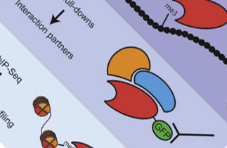
Vermeulen and his colleagues applied quantitative mass spectrometry-based proteomics to identify a large number of novel chromatin “readers” that interact with methylated histone tails. The authors then applied GFP-tag based protein affinity purifications, ChIP-sequencing and conventional protein biochemistry to further validate and functionally characterise their findings. By and large, the known biology of the lysine methylation sites they studied is reflected by their readers, indicating that reader complexes at least partially determine the biological function of the methylation sites they bind to.


In 2009, Michiel Vermeulen received an NWO VIDI grant and a KWF project grant for his research.


Publication:

Quantitative interaction proteomics and genome-wide profiling of epigenetic histone marks and their readers. Michiel Vermeulen, H. Christian Eberl, Filomena Matarese, Hendrik Marks, Sergei Denissov, Falk Butter, Kenneth K. Lee, Jesper V. Olsen, Anthony A. Hyman, Henk G. Stunnenberg and Matthias Mann.