Fighting the cause of Chagas disease

Reducing the parasite population with a virus

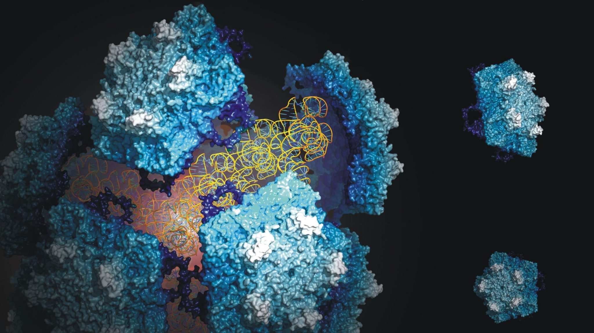
The hitherto fairly unknown Triatoma virus, which kills insects that spread the tropical disease known as Chagas disease, could be used as a pesticide to control the insect plague and thus the spread of the disease. Researchers of the Biomolecular Mass Spectrometry and Proteomics Group in collaboration with researchers of VU University Amsterdam have studied every little detail of the virus and published their findings on 28 April in Nature Chemistry.

 According to assessments of the World Health Organisation, ten million people had Chagas disease in 2012. At least 20,000 people die from the disease every year. This tropical disease is transferred by parasites in blood-sucking insects. The disease can be treated in the first few months, but when a longer period of time has elapsed, patients can no longer be saved. The disease is prevalent in Latin America, but has meanwhile spread to the north and infections have been found in the North-American state of Florida.

Fighting the cause
One of the best ways to prevent the spread of the disease is to combat the insect population that carries the pathogenic parasite. The Triatoma virus offers options in this respect, because infection with the virus kills the insects. In an attempt to chart this virus, researchers of the group of Professor Albert Heck (Utrecht University) in cooperation with the group of Dr Wouter Roos (VU University Amsterdam) have studied the composition, structure and stability of the virus in detail with the help of mass spectrometry and atomic force microscopy.

A complex collaboration
The researchers have shown that the stability of the virus is determined by a complex collaboration between the virus and its genetic material, which is strongly influenced by environmental factors. The experiments also provided information about the structure of the virus and the functioning of the genome. Both processes are vital to the virus’ life cycle. Furthermore, the data contributes to the design of viral systems in nano-technology and medicine.

Structure and stability
Albert Heck: “Our work has provided a lot of insight into the structure and stability of the virus. We have used many techniques in order to find out how to extract the genome from the virus and to reassemble the virus afterwards. This offers opportunities to add new elements to the virus which are potentially lethal to the host.”


This research was sponsored by the Netherlands Proteomics Centre, the Netherlands Organisation for Scientific Research (NWO) and the Foundation for Fundamental Research on Matter (FOM)

Publication
J. Snijder, C. Uetrecht, R.J. Rose, R. Sanchez-Eugenia, G.A. Marti, J. Agirre, D.M.A. Guerin, G.J.L. Wuite, A.J.R. Heck, W.H. Roos
Probing the biophysical interplay between a viral genome and its capsid.
Nature Chemistry
 

Top publication: Deletion of genes causes cancer

In a recent Nature paper researchers from the Hubrecht Institute in cooperation with researchers from the University Medical Center Utrecht (UMC Utrecht), University Utrecht (UU) and the Netherlands Proteomics Centre (NPC), describe a gene that limits the growth of intestinal adenomas. The research might be a lead in the treatment of intestinal cancer. Stem cells in the gut continuously provide new tissue. Prof. Hans Clevers succeeded earlier in identifying and isolating these stem cells.

Together with Dr. Madelon Maurice of the UMC Utrecht and Prof. Albert Heck of the UU and the NPC, the Clevers group searched for genes which are only active in the intestinal stem cells. They found RNF43. When this gene is deleted, exponential growth of the intestinal stem cells cause adenomas, a pre stage of intestinal cancer.

LGR5+ stem cells reside at crypt bottoms, intermingled with Paneth cells that provide Wnt, Notch and epidermal growth factor signals. In this article the researchers find that the related RNF43 and ZNRF3 transmembrane E3 ubiquitin ligases are uniquely expressed in LGR5+ stem cells. Simultaneous deletion of the two genes encoding these proteins in the intestinal epithelium of mice induces rapidly growing adenomas containing high numbers of Paneth and LGR5+ stem cells. In vitro, growth of organoids derived from these adenomas is arrested when Wnt secretion is inhibited, indicating a dependence of the adenoma stem cells on Wnt produced by adenoma Paneth cells. In the HEK293T human cancer cell line, expression of RNF43 blocks Wnt responses and targets surface-expressed frizzled receptors to lysosomes. In the RNF43-mutant colorectal cancer cell line HCT116, reconstitution of RNF43 expression removes its response to exogenous Wnt. We conclude that RNF43 and ZNRF3 reduce Wnt signals by selectively ubiquitinating frizzled receptors, thereby targeting these Wnt receptors for degradation.

Grant for Maarten Altelaar

Utrecht University (UU) has awarded Maarten Altelaar (Biomolecular Mass Spectrometry & Proteomics Group and NPC Hotel Manager) a grant of 250k euros. Altelaar received the grant within the context of the European Research Council (ERC) incentive programme of the Executive Board of Utrecht University. In total four researchers received this grant, whose research proposal has been marked eligible by the ERC.

Network Medicine: the pertubed cancer proteome
Unraveling the effect of cellular perturbations on proteome wide scale is extremely challenging as the proteome is a highly dynamic entity compared to the relative static genome, with protein expression, degradation, posttranslational modification and localization being highly divergent in time. Additional complexity is created by the interconnectivity of the proteins and protein networks making up the functional proteome, requiring high throughput techniques such as quantitative mass spectrometry to study biological/molecular mechanisms in full. In this project we will develop novel proteomics technology to address this complexity and use melanoma as model system. Melanoma shows a rapid increase in incidence and is known for its aggressive nature in development of malignant metastasis and therapeutic resistance. The oncogenic BRAF(V600E) protein kinase, which leads to activation of the RAS–RAF–MEK–ERK signaling pathway, is frequently mutated in human cancer, including approximately 70% of malignant melanomas. In on-going collaborative efforts with the NKI we target the BRAFV600E proteome using high throughput (phospho)proteomics and the current grant will be assigned to the extraction of biologically relevance from the generated proteomics data by developing novel bioinformatics tools.

Top Publication: Genes & proteins involved in the regeneration of the small intestine uncovered

Genes & proteins involved in the regeneration of the small intestine uncovered

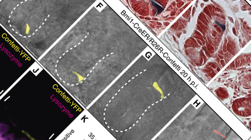
In a collaboration involving several partners in the Utrecht Life Sciences Initiative (ULS) members of the Netherlands Proteomics Centre (Heck group) and the Hubrecht Institute (Clevers group, Alexander van Oudenaarden group, presently still at the Department of Physics, MIT, Cambridge, USA) made a significant advance in understanding the molecular basis that program the regeneration of tissue through the stem cells in the intestine. Joint efforts from these groups allowed the identification of genes and proteins that are specifically expressed in the stem cells of the intestine and that allow this tissue to regenerate. These results have been jointly published in The EMBO Journal on June 12, 2012.

Stem cells receive special attention in the scientific community and in society. This has been inspired by their potential to differentiate into tissues and there use in regenerative medicine. A lot of progress has been made towards the application of stem cells in therapy. Hans Clevers explains: “with our current knowledge it is possible to isolate these stem cells from the intestine and generate organoids in the lab that quite resemble the tissue”. To achieve successfully cellular replacement therapies it is still far from the bedside: “All future applications of stem cells will depend on exquisite understanding of the mechanisms that regulate stem cell biology” ads Clevers. Traditionally, research on stem cells has been focused on the study of a small group of genes, one of them, Lgr5, led Clevers and co-workers to the identification of the stem cells in the intestine and other tissues. However, during the last decade emerging technologies, such as proteomics, have allowed scientists to study thousands of players in the reprogramming process simultaneously. “Our research focused on the identification, in a global fashion, of genes and proteins that are uniquely expressed in the intestinal stem cells and how they behave when stem cells differentiate” says Albert Heck.  In this report, 510 genes were identified to be uniquely expressed in these cells, including Lgr5, defining a stem cell signature. “This is an important first step, providing a clear picture of the molecular signature that provides these cells with their unique properties” explains Javier Munoz, first author on the paper.

13,5 million euro for large-scale research facility coordinated by Albert Heck

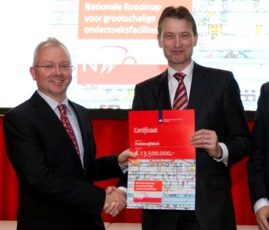
A programme coordinated by Albert Heck, scientific director of the Netherlands Proteomics Centre, has received financing from NWO to set up a large-scale research facility. The project ‘Proteins@Work’, in which various Dutch research institutes collaborate, receives 13,5 million euro. This was announced by State Secretary Zijlstra of OCW and Jos Engelen, chairman of NWO, this afternoon in Utrecht. The participating partners will make state-of-the-art technology, equipment and expertise in the field of the life sciences available to other researchers in the Netherlands.

In total, five programmes in the Netherlands receive financing from NWO within the National Road Map for Large-scale Research Facilities, for which 80 million euro was made available.

Proteins@Work

The Proteins@Work-facility will make high-level technology, equipment and expertise for studying proteins in cells and tissues available to the biological and biomedical researchers in the Netherlands. The facility is of great benefit to Dutch Life Sciences and, in particular, will aid in the prevention and diagnosis of disease. “We work with everybody who wants to understand how proteins work, for example how proteins can cause disease, but also how stem cells can develop into healthy organs”, says Albert Heck, the initiator of Proteins@Work. “Proteins are the essential workhorses in cells and to understand life, we need to know how proteins work together.” The Proteins@Work programme is a collaboration between Utrecht University, the UMC Utrecht, the Hubrecht Institute, the Erasmus MC Rotterdam and the Netherlands Cancer Institute and builds upon the Netherlands Proteomics Centre and the European large-scale proteomics facility, PRIME-XS.

More information

http://www.proteinsatwork.nl/


Martje Ebberink, communications NPC, (030) 253 4564, ebberink@npc.genomics.nl.

NPC on RTL 4 on September 25

Sunday 4 September marks the start of the six-part series Lang Zullen We Leven on RTL 4. In this non-specialist series on DNA and proteomics presenter Geert Hoes investigates the latest developments in the field of DNA and proteomics. The 4th episode on 25 September will focus on proteomics and has been developed in close collaboration with the Netherlands Proteomics Centre.

Channel: RTL 4 ….

Date: September 25, 2011

Time: 11.25

Episode: 4

Dell promotional clip

In February this year our group participated in a promotional video clip for Dell in which they present their recently obtained data storage cluster for proteomics data.

When a fluorescent sea anemone glows on a coral reef, it’s using a protein. When we move, proteins help our muscles contract. And when we curse our wrinkles as we age, it’s the lack of the protein collagen we lament.

{youtube}tRfN5Os79P4{/youtube}

Proteins also tell us about human health. By identifying proteins linked with specific diseases, we can develop novel diagnostics and therapies. At Utrecht University in the Netherlands, the Mass Spectrometry and Proteomics Group is exploring new methods of protein research. They can identify a protein or peptide every 100 milliseconds, producing vast amounts of data that must be stored safely and easily retrieved. Often, of all the proteins defined for each project, a tiny number are significant. Quick access to data is crucial for efficient analysis and isolation of the proteins that can fuel drug discovery.

The group wanted a new storage solution that would prevent bottlenecks and cut out painstaking backups. The Dell DX Object Storage Platform emerged as the best fit, based on its simplicity, flexibility and integrated data protection. Instead of slow tape backups, the IT team now relies on mirroring and automatic replication. When the group looks after data for other organisations, it knows it’ll be safe and easy to access. And, crucially, researchers can download files twice as quickly because the platform has higher throughput. Without delayed downloads, all their time is dedicated to the discovery of patterns and anomalies in protein behaviour that can change the way we manage disease.

Watch the video to hear the Utrecht University team talk about the challenges and rewards of proteomics.