PhosphoPath: Visualization of Phosphosite-centric Dynamics in Temporal Molecular Networks

PhosphoPath: Cytoscape compatible visualization tool for quantitative PTM-centric proteomics data published

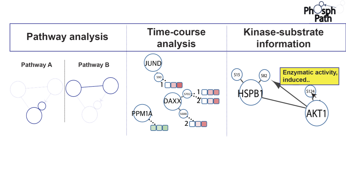
Protein phosphorylation is an essential post-translational modification (PTM) regulating many biological processes at the cellular and multicellular level. Continuous improvements in phosphoproteomics technology allow the analysis of this PTM in an expanding biological content. Yet, up till now proteome data visualization tools are still very gene centric, hampering the ability to comprehensively map and study PTM dynamics. Here we present PhosphoPath, a Cytoscape app designed for the visualization and analysis of quantitative proteome and phosphoproteome datasets. PhosphoPath brings knowledge into the biological network by importing publically available data, and enables PTM site-specific visualization of information from quantitative time series. To showcase PhosphoPath performance we use a quantitative proteomics dataset comparing patient derived melanoma cell lines grown either in conventional cell culture or xenografts.

PhosphoPath: Visualization of phosphosite-centric dynamics in temporal molecular networks

50 free e-prints of the final published article are available for interested colleagues: click here


Prestigious American Chemical Society award for Albert Heck

The American Chemical Society has honoured Albert Heck with the ‘ACS Frank H. Field and Joe L. Franklin Award for Outstanding Achievements in Mass Spectrometry’. Heck receives the award for his development of new methods and techniques to identify and study the structure and function of proteins and protein complexes.

In the field of Chemistry, the American Chemical Society Awards are considered to be very prestigious, and are almost exclusively presented to researchers from America. The Award will be presented during the meetings of the American Chemical Society in March.

Publication in Cell Reports

Last year two first drafts of the human proteome were made available [1,2]. These drafts provide an overview of all proteins detected in different cells and tissues of our body. As proteins are the prime molecular entities responsible for all biological processes these drafts tell us which “players” are present. A next step in the analysis of the proteome is to observe how proteins work and communicate with each other. They most often do this by the transfer of a chemical phosphate moiety from one to the other protein, which may lead to activation or deactivation of the protein modified.

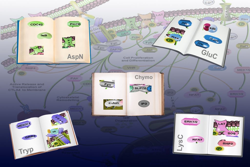
Proteomics researchers have been aiming at unraveling this communication language, termed the phosphoproteome. But as Piero Giansanti et al. [3] show in the latest issue of Cell Reports, these researchers have been using a single dictionary (i.e. workflow) for that, which can only help to translate a specific part of the communication. By using 5 different dictionaries (5 different workflows) Giansanti et al. are able to provide a much broader view into cellular communication, and show indeed that in the public repositories there is a considerable bias in the phosphoproteome. Each dictionary was able to translate about 70% new parts of communication, whereas only 7% of what the cells were communicating could be translated by all 5 different workflows. More technically, the 5 “dictionaries” are presented by 5 different proteases that were used to cleave the proteins into for analysis amendable peptide sequence reads. Using this multiple protease they clearly showed that optimal detection of each protein individual phosphorylation event is linked to a preferred protease.

In the proteolysis step the greatest loss occurs as many of the generated small peptides, get lost in translation. Giansanti et al. repair this problem and present an augmented human phosphopeptide atlas of 37,771 unique phosphopeptides, each representing a “word of communication” in the cell.

1. Kim, M. S., Pinto, S. M., …. & Pandey, A. (2014) A draft map of the human proteome, Nature. 509, 575-81.
2. Wilhelm, M., Schlegl, J., ….. & Kuster, B. (2014) Mass-spectrometry-based draft of the human proteome, Nature. 509, 582-7.
3. Giansanti, P, Aye, T.T., van den Toorn, H., Peng, M., van Breukelen, B. & Heck A.J.R (2015) An augmented multiple protease based human phosphopeptide atlas, Cell Reports

You can listen to the radio interview with Albert Heck about this research project here

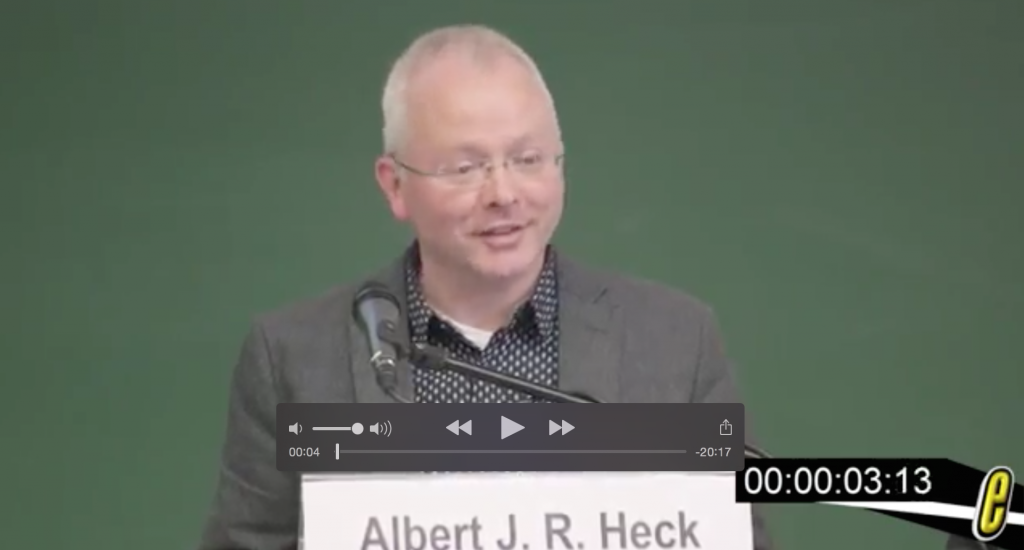
Seminars by Albert Heck at the Proteomics forum

Prof Albert Heck gave an interesting seminar at the Proteomics forum in Berlin (march 2015). You can view these seminars by following the youtube links below.

Albert Heck Seminar part 1: Preview:

https://www.youtube.com/watch?v=S1uaJ9GZ420

Albert Heck Seminar part 2: Preview:

https://www.youtube.com/watch?v=oD5gNi7_h38

First article published using the new Fusion MS

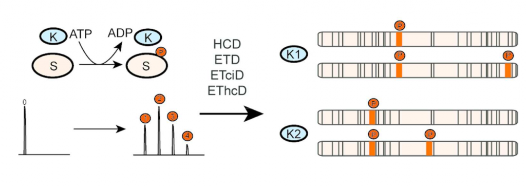
Top-down analysis of intact proteins by mass spectrometry provides an ideal platform for comprehensive proteoform characterization, in particular, for the identification and localization of post-translational modifications (PTM) co-occurring on a protein. One of the main bottlenecks in top-down proteomics is insufficient protein sequence coverage caused by incomplete protein fragmentation. Based on previous work on peptides, increasing sequence coverage and PTM localization by combining sequential ETD and HCD fragmentation in a single fragmentation event, we hypothesized that protein sequence coverage and phospho-proteoform characterization could be equally improved by this new dual fragmentation method termed EThcD, recently been made available on the Orbitrap Fusion. Here, we systematically benchmark the performance of several (hybrid) fragmentation methods for intact protein analysis on an Orbitrap Fusion, using as a model system a 17.5 kDa N-terminal fragment of the mitotic regulator Bora. During cell division Bora becomes multiply phosphorylated by a variety of cell cycle kinases, including Aurora A and Plk1, albeit at distinctive sites. Here, we monitor the phosphorylation of Bora by Aurora A and Plk1, analyzing the generated distinctive phospho-proteoforms by top-down fragmentation. We show that EThcD and ETciD on a Fusion are feasible and capable of providing richer fragmentation spectra compared to HCD or ETD alone, increasing protein sequence coverage, and thereby facilitating phosphosite localization and the determination of kinase specific phosphorylation sites in these phospho-proteoforms. Data are available via ProteomeXchange with identifier PXD001845.

Integrated OMICS approach reveals ROCK1 kinase as potential combinatorial drug target in BRAF mutant melanoma

In a recent, open access, article in Molecular Systems Biology researchers from the Biomolecular Mass Spectrometry and Proteomics Group at Utrecht University and The Netherlands Cancer Institute in Amsterdam, report on an integrated analysis of proteomic and phospho‐proteomic data from BRAF inhibitor‐treated melanoma cells and a functional genomic screen for shRNAs sensitizing melanoma to BRAF inhibitor treatment, which identifies ROCK1 kinase as a combinatorial drug target.

Treatment of BRAF mutant melanomas with specific BRAF inhibitors leads to tumor remission. However, most patients eventually relapse due to drug resistance. The researchers designed an integrated strategy using (phospho)proteomic and functional genomic platforms to identify drug targets whose inhibition sensitizes melanoma cells to BRAF inhibition. They found many proteins to be induced upon PLX4720 (BRAF inhibitor) treatment that are known to be involved in BRAF inhibitor resistance, including FOXD3 and ErbB3. Several proteins were down‐regulated, including Rnd3, a negative regulator of ROCK1 kinase.

For the genomic approach, they performed two parallel shRNA screens using a kinome library to identify genes whose inhibition sensitizes to BRAF or ERK inhibitor treatment. By integrating the functional genomic and (phospho)proteomic data, they identified ROCK1 as a potential drug target for BRAF mutant melanoma. ROCK1 silencing increased melanoma cell elimination when combined with BRAF or ERK inhibitor treatment.

Translating this to a preclinical setting, a ROCK inhibitor showed augmented melanoma cell death upon BRAF or ERK inhibition in vitro. This research collaboration provides data that merits exploration of ROCK1 as a target in combination with current BRAF mutant melanoma therapies.

Article
ROCK1 is a potential combinatorial drug target for BRAF mutant melanoma
Marjon A Smit, Gianluca Maddalo, Kylie Greig, Linsey M Raaijmakers, Patricia A Possik, Bas van Breukelen, Salvatore Cappadona, Albert JR Heck, AF Maarten Altelaar, Daniel S Peeper
Molecular Systems Biology(2014)10:772; DOI 10.15252/msb.20145450

Funding
This work was funded by the NWO VIDI grant (723.012.102) awarded to Maarten Altelaar, an International postdoc grant from Vetenskapsrådet (VR), Sweden awarded to Gianluca Maddalo, by the Netherlands Proteomics Center (NPC), the Netherlands Organization for Scientific Research (NWO) funded large-scale proteomics facility Proteins At Work (project 184.032.201), by the European Community’s Seventh Framework Programme (FP7/2007–2013) for the PRIME-XS project grant agreement number 262067, a Queen Wilhelmina award and a grant from KWF Kankerbestrijding


Unprecedented cooperation cracks the programming of the cell

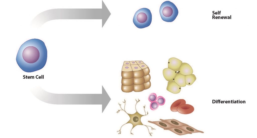
An international consortium including Prof. Albert Heck and former lab members Javier Munoz and Marco Benevento, has uncovered the molecular events underlying the biological processes of the creation of stem cells from specialized (e.g. skin) cells, a process called cellular reprogramming. The team also discovered a new type of stem cells termed F-class that have unique properties compared to the previously known stem cell types. This opens up new avenues for generating useful “designer” cells, which might not necessarily exist in the body or during development but could be safer and more efficient when used for drug evaluation in personalized therapy. The importance and anticipated impact of this research is supported by the unprecedented coordinated publication of 5 scientific articles in Nature and Nature Communications on December 10, 2014.

The research project involved a collaboration of 27 expert key researchers in  stem cell biology, proteomics, epigenetics and RNA biology. ‘This is the first time an integrative biology study was carried out so comprehensively. Our team has catalogued every major biological checkpoint of the reprogramming process, identifying which combination of genes and proteins, and modifications thereof, are associated with each step. By doing this, I truly believe you can say we cracked the programming of the cell’, says Dr. Albert Heck, Professor of Biomolecular Mass Spectrometry and Proteomics at Utrecht University.

More economically
The extremely in depth analysis of the process of reprogramming specialized cells into stem cells focused on learning how to control the paths to either the new F-class stem cell versus “traditional”, embryonic-like stem cells. Comparing the two cell types revealed that the new class of stem cells is easier, less expensive and faster to grow compared with the embryonic-like stem cells. Because of these properties, the new “F-class” stem cells can be produced more economically in very large quantities, which will speed up drug screening efforts, disease modelling and eventually the development of treatments for different illnesses.

Clinical Implications
Stem cells hold promise for future medicine aiming to treat and cure currently incurable diseases such as blindness, Parkinson’s, Alzheimer’s, spinal cord injury, stroke, diabetes, blood and kidney diseases, which are associated with tissue damage and cell loss. The uncovered detailed knowledge of cellular reprogramming will help to better understand this process, which is critical to generating safe and highly efficient sources for therapeutic cell production.

Prinicipal investigators

The principal investigators of this project are:

  • Cellular reprogramming & and overall project co-ordination: Dr. Andras Nagy, the Canada Research Chair in Stem Cells and Regeneration at the Lunenfeld-Tanenbaum Research Institute, of the University of Toronto, Canada. Nagy established Canada’s first human embryonic stem cell lines and also discovered a new method to create induced pluripotent stem cells.
  • Epigenetics: Dr. Jeong-Sun Seo, Professor in the Department of Biochemistry at Seoul National University, College of Medicine, South Korea
  • Proteomics: Dr. Albert Heck, Professor of Biomolecular Mass Spectrometry and Proteomics at Utrecht University, the Netherlands. Since 2003, he has also been the scientific director of the Netherlands Proteomics Centre. Dr. Heck is well known for the development of innovative enabling technologies in proteomics and was awarded the HUPO Discovery Award in 2013 and EuPA’s Proteomics Pioneer Award 2014.
  • RNA biology: Dr. Thomas Preiss, Professor of RNA Biology at The John Curtin School of Medical Research at The Australian National University in Canberra. Dr Preiss is well known for his research into the patterns and mechanisms of gene regulation at the RNA level with an international profile as one of Australia’s foremost experts in this area.

This research was funded by many sources, but in the Netherlands specifically by the Netherlands Proteomics Centre, by the Netherlands Organization for Scientific Research (NWO) funded large-scale proteomics facility Proteins At Work (project 184.032.201) and by the European Community’s Seventh Framework Programme (FP7/2007–2013) for the PRIME-XS project grant agreement number 262067.